Transesophageal Echocardiography (TEE, TOE, Endoscopic ECHO)
Similar to endoscopy, a probe approximately 90-100 cm in length is swallowed after numbing the throat and administering a sedative through a vein.
Images taken from this probe, unlike those obtained from external body examinations, offer very clear visuals because there are no significant muscle and bone structures in front and the imaging is done very close to the heart.
Even the smallest details are captured, including color flows and Doppler examinations. This method is not performed in every patient.
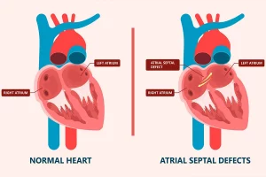
It is more commonly used in cases where there is a possibility of clots in the atria and ventricles (such as after a stroke, etc.), in patients who have undergone surgical replacement of the mitral and aortic valves for the evaluation of artificial valve function, in the examination of the substructures of the mitral valve, in aortic ruptures, in inflammations of the valves, and in the definitive diagnosis of interatrial and interventricular defects.
Why Is TEE Performed?
Doctors use TEE when they need a closer look at the heart than a regular echocardiogram can provide. It’s helpful for detecting blood clots, heart valve problems, infections, or issues with the heart’s upper chambers.
It’s often used before surgery or procedures like valve repair, or when other tests didn’t give enough information.
What to Expect During the Procedure
Before the test, your throat will be numbed, and you may get a mild sedative to help you relax. The doctor then gently guides the probe down your throat. The whole procedure usually takes 30 to 60 minutes.
After the procedure, you’ll need to rest until the sedative wears off, and you shouldn’t eat or drink for a short time to allow your throat to recover.