Computed Tomographic Angiography (Coronary CT/Virtual Angiography)
Computed tomographic angiography, also known as CT angiography or CTA, is a method that combines traditional computed tomography with angiography, providing detailed information about the blood vessels in our body.
In a classic computed tomography (CT) scan, cross-sectional or sliced images of the body are obtained. In the resulting image, including all organs and tissues, the slices are segmented.
As these slices are cut at specific intervals, for example, at intervals of 1-5 mm, dozens of slices are obtained when evaluating a particular region thoroughly. In these images, the blood vessels appear as small structures. Computer systems combine these slices to create three-dimensional images.
If specifically the arteries are to be visualized, during the procedure, the flow of a dye (contrast agent) from a vein is tracked within the blood vessels, and after the slices are combined, a three-dimensional vascular film, namely an angiography, is obtained, which may include only the vessels or vessels with another tissue, such as bones.
If the examination is for the veins, the procedure is called CT venography. During CT angiography, while the agent is given from any vein in the arm, when the dye mixes into the arterial system, arterial imaging, i.e., CT angiography, is obtained.
In contrast, for classic arterial angiography, a catheter must be inserted into the artery to capture the film. Therefore, CT angiography is considered less invasive or less discomforting as there is no catheter involved.
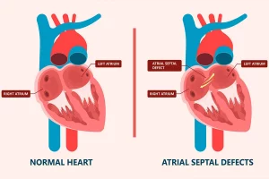
CT angiography is a test that can be used to diagnose constrictions, blockages, aneurysms, deep vein thrombosis, pulmonary embolism, and other vascular diseases. With CT angiography, narrowings and constrictions in the blood vessels are evaluated more accurately and in more detail compared to magnetic resonance imaging and ultrasound.
In many cases, CT angiography is now performed instead of conventional angiography because it does not require a catheter, causing less discomfort and providing accurate information about the vessels.
Due to its ability to provide accurate information about the vessels and its less invasive nature, CT angiography can also be used as a screening test. There is radiation exposure during CT angiography, but there is no radiation risk after the examination is completed.
Preparation
Fasting is required at least 4 hours before the examination. However, if the patient is taking any medications, they should continue taking them. During the examination, clothing and accessories, including metal objects, are removed.
If there is a history of kidney disease, heart disease, or thyroid disease, the doctor should be informed. This examination should not be performed if the patient is pregnant or suspected to be pregnant.
Risks
CT angiography carries a risk of radiation as it is an examination performed using X-rays.
The contrast agent used during the examination can cause allergic reactions. If allergic reactions have occurred to iodine-containing substances or contrast agents used in similar examinations in the past, this procedure should be avoided.
Additionally, the contrast agent can further impair the kidneys as it is excreted from the body through them. Patients with kidney problems should inform their doctor before this examination. Blood tests can provide information about the condition of the kidneys and whether CT angiography can be performed.
CT angiography should not be performed in the following cases:
- If there is an allergy to the contrast agent
- If there are kidney problems
- In the case of severe diabetes
- During pregnancy or suspected pregnancy
- In extremely obese patients
- In patients with very poor general condition and body functions.
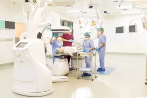
How to do it?
During the examination, the patient lies on a large table that moves into a wheel-shaped device. Meanwhile, a contrast agent (dye) is injected into one of the veins in the arm. The injection of the contrast agent may cause a temporary burning sensation, pain, or nausea in the arm.
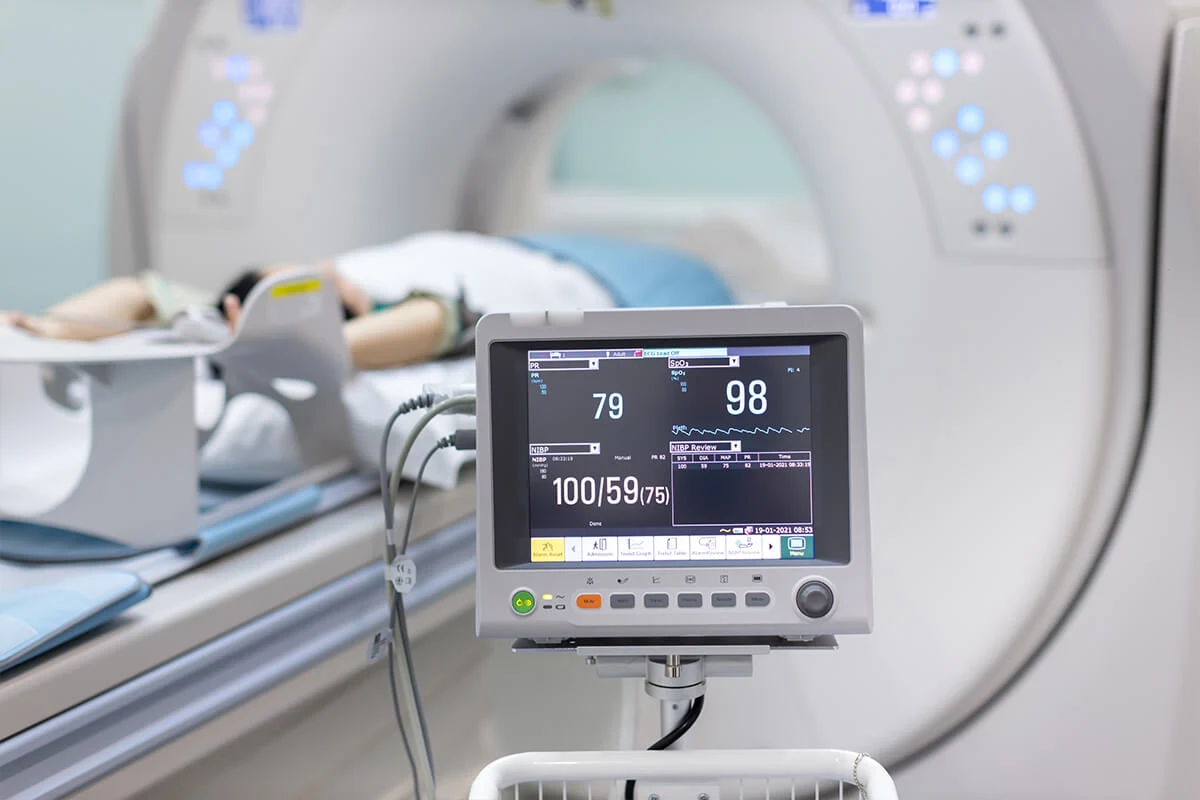
The patient should remain still throughout the examination. The technician may sometimes ask the patient to hold their breath for 10-25 seconds. The table automatically moves into the device, and during this time, sectional images of the area to be examined are taken.
Afterwards, the computer generates three-dimensional images. The entire examination typically takes 20-60 minutes.
What can happen after CT angiography?
After a CT angiography, patients can continue their daily activities without any restrictions. However, they are encouraged to consume plenty of water or fluids to help eliminate the contrast agent from their system as quickly as possible.
Side effects?
CT angiography’s most significant early side effect is allergic reactions to the contrast agent. These reactions typically occur immediately and may manifest as redness, itching, and in rare cases, difficulty breathing and swallowing.
If the contrast agent leaks from the injection site into the skin, it can cause redness, pain, and swelling. The most important side effect of the contrast agent is its potential to exacerbate kidney function, especially in patients with mild kidney impairment.
What are the shortcomings of CT angiography?
- Excessively obese patients may not fit inside the device for the examination.
- CT angiography cannot be performed in patients with kidney disease as the contrast agent may further impair kidney function.
- When the heart cannot pump blood adequately or when there is insufficient blood flow in the vessels, the evaluation may not be highly reliable.