COLOR DOPPLER ECOCARDIOGRAPHY (ECO HEART ULTASOUND)
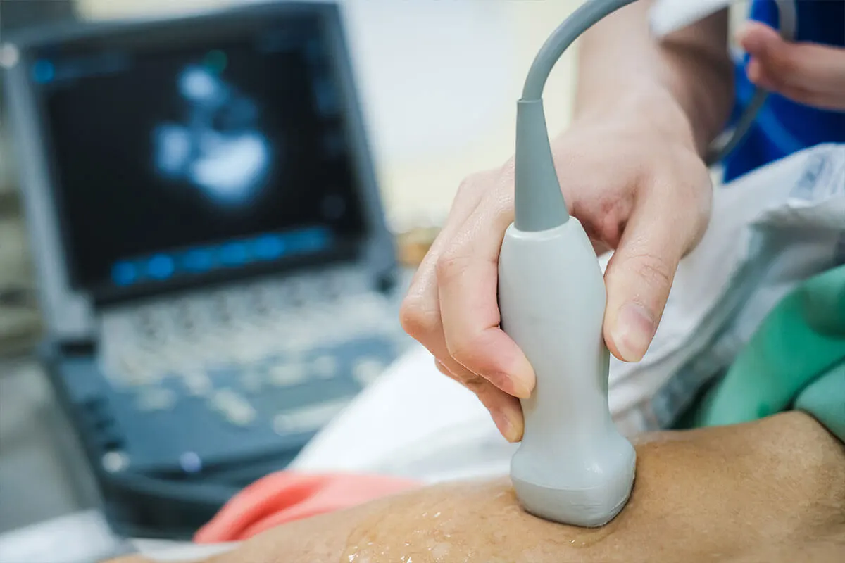
Sending ultrasound waves to the heart from the front of the chest with a device and converting the received waves into an image on the device is known as echocardiography. With this examination:
- Evaluation of the heart chambers, the outgoing aortic vessel, and the heart membrane
- Assessment of heart wall thickness, heart contraction function (ejection fraction – EF)
- Heart valve functions, stenosis, insufficiencies, and infections
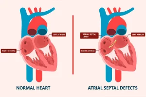
- Congenital (birth) heart diseases and heart defects can be evaluated.
The advantage of this method is that it can be performed at any center, it is inexpensive, harmless, and a radiation-free method.
Why Is It Done?
Doctors use echocardiography to check for many heart conditions. It can show how your heart valves are working, if there’s fluid around the heart, or if the heart muscle is weak. It’s also used after a heart attack or when a patient has chest pain, shortness of breath, or irregular heartbeat.
What to Expect During the Test
During the test, you lie on a table while a technician moves a small device called a transducer over your chest. The device sends sound waves that create moving images of your heart. The test usually takes 30 to 60 minutes, and you can go back to your normal activities right after it’s done.